
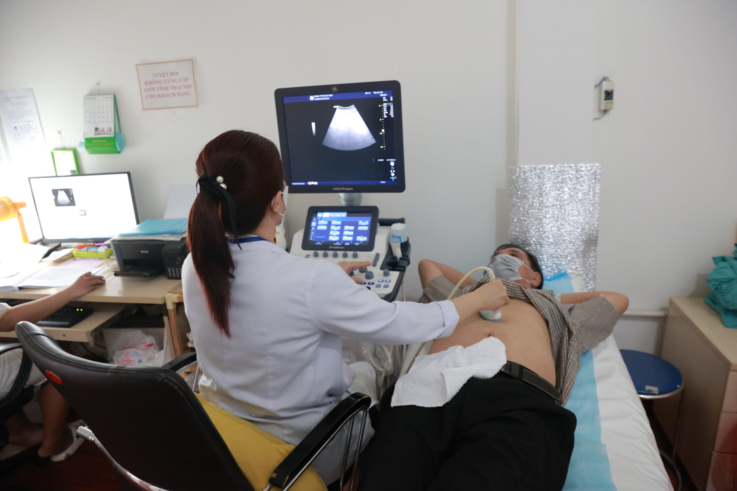
An abdominal ultrasound uses high-frequency sound waves to create real-time images of organs inside the abdomen. The technique involves no X-rays or radiation, causes no pain, and is completely safe — including during pregnancy. A single scan can assess multiple organs simultaneously: the liver, gallbladder, bile ducts, pancreas, spleen, kidneys, bladder, and, in women, the uterus and ovaries. It is one of the most versatile and widely used diagnostic tools in medicine.
When Is an Abdominal Ultrasound Recommended?
Doctors commonly order this scan when patients experience:
- Unexplained or recurrent abdominal pain in any part of the abdomen.
- Liver and gallbladder symptoms: Jaundice (yellowing of the skin or eyes), pain in the upper right abdomen, or suspected gallstones.
- Liver disease monitoring: Following up on hepatitis, cirrhosis, or fatty liver disease.
- Suspected kidney problems: Back or flank pain, blood in the urine, painful urination, or possible kidney stones.
- Routine health screening: Detecting abnormalities in abdominal organs before symptoms develop.
- Masses or lumps: Identifying, characterising, and monitoring abdominal tumours or cysts.
- Post-treatment monitoring: Checking outcomes after abdominal surgery or treatment.
- Emergency assessment: Rapidly evaluating suspected organ rupture, fluid in the abdomen, or abdominal trauma.
How to Prepare for an Abdominal Ultrasound
Proper preparation is essential — particularly for clear views of the gallbladder and pancreas:
Fasting:
- Fast for at least 6 to 8 hours before the scan — ideally 8 to 12 hours (a morning appointment after fasting overnight is ideal). Food and gas in the intestines can significantly obscure the ultrasound image, especially in the gallbladder and pancreatic areas.
- Small sips of plain water are generally permitted if needed to take essential daily medications.
Full bladder (when required):
- If the scan includes the bladder, uterus, or ovaries, your doctor may ask you to drink four to six glasses of water and avoid urinating for approximately one hour beforehand. A full bladder acts as a window that allows sound waves to pass through more clearly and improve visibility of structures behind it.
Clothing:
- Wear comfortable, loose-fitting clothes that can be easily pulled up to expose the abdomen.
Medications:
- Continue taking your usual daily medications with a small amount of water unless specifically instructed otherwise by your doctor.
What Happens During the Scan?
- You lie on your back on the examination table. The technologist or doctor may ask you to roll onto your side to obtain better views of certain organs.
- A clear, water-based gel is applied to your skin. The gel helps transmit sound waves and does not stain or irritate.
- A handheld probe is moved gently across the skin of your abdomen in various directions and angles to capture images of all relevant structures.
- The procedure typically takes 15 to 30 minutes.
- Once finished, the gel is wiped away and you can resume normal activities immediately.
What Conditions Can an Abdominal Ultrasound Detect?
- Gallstones, kidney stones, and bile duct stones.
- Fatty liver, hepatitis, cirrhosis, and liver tumours.
- Pancreatitis and pancreatic masses.
- Kidney cysts, hydronephrosis (fluid build-up in the kidney), and kidney tumours.
- Ovarian cysts and uterine fibroids.
- Enlarged spleen.
- Fluid in the abdominal cavity (ascites).
An abdominal ultrasound is one of the fastest, safest, and most cost-effective diagnostic tools available. At Ky Hoa Medical Center, our imaging department is equipped with high-resolution ultrasound machines and staffed by experienced radiologists. Results are available promptly, allowing your doctor to advise on next steps without delay. Schedule your scan today as part of a proactive approach to protecting your health.